HUNTSVILLE – Low-intensity ultrasound therapies may one day rebuild stronger knees following injury or surgery, thanks to research by Dr. Anu Subramanian at the University of
Alabama in Huntsville that’s being supported by the
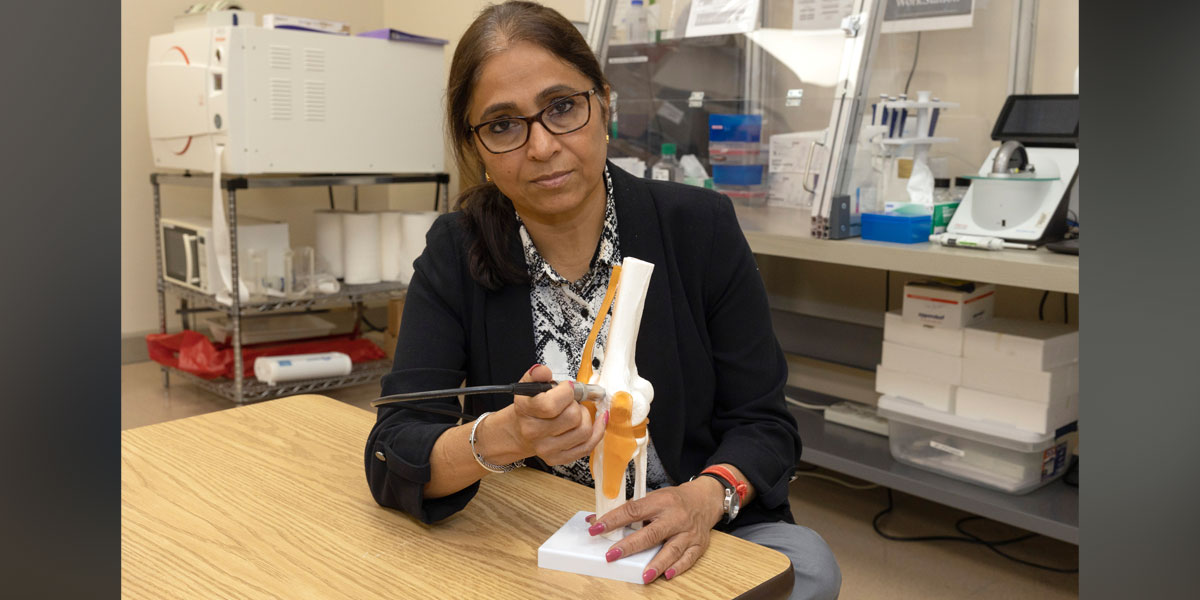
A professor of chemical and materials engineering at UAH, Subramanian has been the principal investigator in research exploring the effects of ultrasound on cartilage regrowth since before coming to UAH in 2018. Her research into in-vitro and bovine cadaver knee research is supported by the National Institutes of Health and Subramanian has been awarded nearly $2 million in grants for her work.
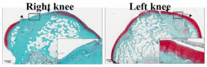
respectively. The solid arrows show the boundaries of the defect. Left rabbit knee joints
were treated with cLIUS and the right knee joints served as the control. A robust red color
between the arrows in the left joint indicates tissue regeneration. (Courtesy Anu Subramanian)
Subramanian’s research has progressed over the years from the test-tube phase through cadaver cow knee work, rabbit testing and now to equine knee investigations in collaboration with Colorado State University.
Cartilage that serves as a cushion between knee bones is a protein-rich matrix with very few cells. It has no blood vessels so it cannot regrow itself. Orthopedic surgeons try to make up for that with a technique that drills into the adjoining bone to create microfractures to produce blood rich in stem cells.
Some of the resulting mesenchymal cells convert to another type, chondrocyte cells that are needed to regrow collagen. But some instead become fibroblasts and create a much less sturdy form of collagen.
The result is a weaker bond and the potential for future failure where the injury or surgical intervention took place. Subramanian has been investigating a low-cost method that uses continuous low-intensity ultrasound therapy to regrow quality post-operative cartilage in joints like the knee or shoulder.
Under a current four-year, $494,000 grant that’s part of her total award of $1,957,903, ultrasound will be used following equine surgeries at CSU.
The advancing research is aimed at a time when an injured knee will undergo Magnetic Resonance Imagery and a computer will tell the surgeon where and how best to apply post-operative ultrasound so that resulting cartilage regrowth has characteristics very close to the original material.
“We have demonstrated the proof-of-concept in a rabbit model, and currently we are working to translate the findings to a larger animal model, the equine model, which has joint characteristics close to that of a human,” Subramanian said. “At CSU, Dr. Brad Nelson and Dr. Jeremiah Easley will be performing the surgeries in sheep and testing the ultrasound regimen to repair defects in sheep. They will also help evaluation of the repair using biomechanical and immunological techniques.”
Nelson is an assistant professor of equine surgery at CSU and Easley is the co-director of the CSU Preclinical Surgical Research Laboratory and an assistant professor in the CSU Department of Clinical Sciences.
“I also hope to continue my collaboration with Dr. Hendrik Viljoen, distinguished professor in the Department of Chemical and Biomolecular Engineering at the University of Nebraska,” she said. “He and I started this project together (there).”
Dr. Sarma Rani, a UAH associate professor of mechanical and aerospace engineering, is a co-investigator in the current grant which also supports doctoral students Shahid Khan (biotechnology science), Aryana Singh Bhati (biotechnology), Sattik Basu (mechanical and aerospace engineering) and Owen Trippany (mechanical and aerospace engineering; chemical and materials engineering).
Earlier rabbit research demonstrated that ultrasound promotes integration of newly grown tissue with the surrounding native tissue, Subramanian said. That was further proof of concept in the stepped research paradigm used to advance technologies such as ultrasound toward human use.
“Briefly, we surgically created defects in the knee joints of rabbits using a technique called microfracture, where a small defect is created and the cartilage is debrided and tiny holes are drilled into the underlying bone to harness the endogenous supply of mesenchymal stem cells that have healing potential,” she said. “We have demonstrated … that joints that received ultrasound upon microfracture had improved cartilage repair and better repair scores when compared to control joints that did not receive any ultrasound, but underwent microfracture.”
The results also demonstrated that ultrasound can encourage repair in the initial pro-inflammatory joint environment that immediately follows surgery or injury, she said.
“Inflammation during the recovery and regeneration phase plays a critical role in modulating the repair outcome,” Subramaian said. “For example, during the early recovery phase, the joint environment harbors a plethora of cytokines moieties that, if left unchecked, can have a catabolic effect and impede the repair process. So, treatment
modalities need to encourage healing and repair, but also mitigate the catabolic effects of the cytokines present.
“Our work has shown that continuous low intensity ultrasound, when used at the beneficial regimen, promotes repair and regeneration in an inflammatory environment.”